
2 D Echo and Colour Doppler Services

Add Your Heading Text Here
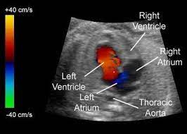
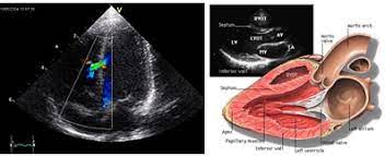
A 2D echocardiogram (2D echo) and color Doppler are specific ultrasound techniques used inpawana specilaity clinic to assess the structure and function of the heart. They provide valuable information about the heart’s chambers, valves, blood flow, and overall cardiac performance. Here are some indications for a 2D echo and color Doppler examination:
Evaluation of Heart Function: A 2D echo and color Doppler can assess the overall function of the heart, including the pumping efficiency, wall motion abnormalities, and ejection fraction (measurement of how well the heart is pumping blood). It helps diagnose conditions like heart failure, cardiomyopathy, or myocardial infarction.
Valvular Heart Disease: These techniques are essential for evaluating the structure and function of heart valves. They can detect abnormalities such as stenosis (narrowing), regurgitation (leakage), or prolapse (bulging) of the valves. 2D echo and color Doppler can also determine the severity of the valve disease and guide further management.
Congenital Heart Disease: 2D echo and color Doppler are crucial in diagnosing and assessing congenital heart defects, which are abnormalities in the heart’s structure present at birth. These techniques help visualize the defects, evaluate blood flow patterns, and monitor the impact on heart function.
Assessment of Cardiac Chambers: The size, shape, and function of the heart chambers can be evaluated using 2D echo and color Doppler. They can detect conditions like enlargement of the heart chambers (cardiomegaly), hypertrophic cardiomyopathy, or chamber wall abnormalities.
Detection of Cardiac Masses or Tumors: 2D echo and color Doppler can help identify and characterize cardiac masses or tumors. They can differentiate between benign and malignant growths and provide information about their location, size, and effects on cardiac function.
Evaluation of Pericardial Effusion: Pericardial effusion refers to the accumulation of fluid in the sac surrounding the heart (pericardium). 2D echo and color Doppler can detect the presence and amount of fluid, assess its impact on heart function, and guide further management, such as pericardiocentesis (fluid drainage).
Assessment of Pulmonary Hypertension: Pulmonary hypertension is high blood pressure in the arteries that carry blood from the heart to the lungs. 2D echo and color Doppler can estimate the pressure in these vessels, evaluate the impact on heart function, and monitor response to treatment.
Preoperative Assessment: 2D echo and color Doppler are often performed as part of the preoperative evaluation before cardiac surgeries. They provide detailed information about the heart’s structure, function, and blood flow, helping the surgical team plan the procedure and anticipate potential complications.
These are some common indications for a 2D echo and color Doppler examination. However, it’s important to note that the specific reasons for performing these tests may vary depending on the patient’s clinical presentation and the cardiologist’s judgment. The tests are typically ordered based on the individual’s symptoms, medical history, physical examination findings, or as part of routine cardiac evaluations.